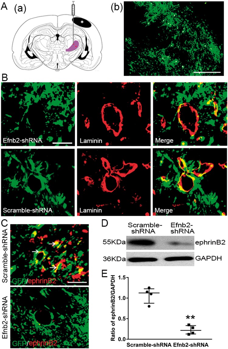
Figure 3.
EphrinB2 knockdown by shRNA in the ipsilateral thalamus after cortical infarction. (Aa) Schematic diagram of brain section (−2.8 mm from bregma) showing the location of cortical infarction (black color) and ipsilateral thalamus (purple color) where lentivirus was injected. (b) GFP-tagged shRNA was considerably detected in the thalamus at seven days after MCAO. (B) Immunoreactivity showing that GFP-tagged Efnb2-shRNA and Scramble-shRNA were expressed in laminin-immunopositive cells in the ipsilateral thalamus at seven days after MCAO. (C) Immunoreactivity showing co-staining of ephrinB2 with GFP-tagged shRNA. Scale bar: 50 µm. (D) EphrinB2 knockdown markedly decreased ephrinB2 levels. (E) Quantitative analysis of ephrinB2 expression. n = 4, data are expressed as median ± interquartile range. **P < 0.01 compared with the Scramble-shRNA group.

