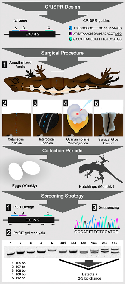
Figure 1. Gene Editing in Lizards through Microinjection of Ovarian Follicles.
Flow diagram detailing CRISPR design, surgical procedure, collection periods, and screening strategy. CRISPR design shows the placement and sequence of CRISPR guides A (blue), B (pink), and C (cyan) within exon 2 of the tyr gene; protospacer adjacent motif (PAM) sites are underlined. Surgical procedure: lizard anesthesia and the surgical steps to access and microinject ovary follicles. Collection periods: the time between gathering eggs and raising hatchlings. Screening strategy: the steps used to detect tyr crispants, including (1) PCR primer design; (2) PAGE analysis, which can reliably detect 2–3 bp changes; and (3) Sanger sequencing.

