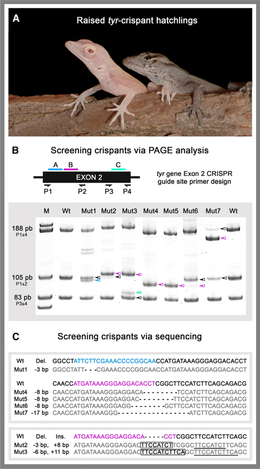
Figure 2. Detection and Sequencing of tyr Crispants.
(A) Albino tyr crispant (left) and wild-type (right) age-matched hatchlings.
(B) PCR primer placement (P1–P4) relative to CRISPR target sites A (blue), B (pink), and C (cyan). Representative PAGE results are shown for seven of the mutant lizards. Colored arrows denote bands with altered mobility relative to wild type (WT).
(C) Sequences of CRISPR-Cas9-induced indels from representative tyr crispants. (Top) Mut1 and Mut4–7 sequences with deletions. (Bottom) Mut2 and Mut3 sequences with insertions. Targeted guide sites A (blue), B (pink), and C (cyan) are highlighted in WT reference sequences. tyr crispants deletions are indicated in bold, insertions boxed, and sequences matching WT in gray.
See also Figure S2.

