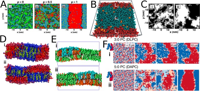
Figure 20.
Factors affecting the nature of phase separation in coarse-grained simulations. (A) Morphologies of domains in DLiPC/PLiPC/DPPC/cholesterol mixtures as a function of DLiPC/(DLiPC + PLiPC) ratio (ρ). The mixtures shown here are those with ρ = (i) 0, (ii) 0.5, and (iii) 1. The membrane is tessellated with areas assigned to cholesterol shown in yellow, DPPC in blue, PLiPC in green, and DLiPC in red. The domain boundaries are shown as black lines. The phase separation becomes stronger with increasing ρ. Adapted with permission from ref (573). Copyright 2015 American Chemical Society. (B) The partitioning of POPC (orange) at the Lo/Ld phase boundary. The liquid-ordered phase is formed mainly by DPPC (cyan) and cholesterol (gray), whereas the liquid-disordered phase consists mainly of DLiPC (red). Adapted with permission from ref (574). Copyright 2010 Elsevier. (C) The effect of transmembrane peptides on the alignment of Lo and Ld phases across leaflets in the DPPC/PLiPC/DLiPC/cholesterol mixture. Aligned Lo phases are shown in white, aligned Ld phases in black, and nonaligned regions in gray. Here, ρ = 0.6 (see panel A). Data are shown for (i) the peptide-free system and (ii) a system with 4 mol % WALP-23. Adapted with permission from ref (575). Copyright 2016 American Chemical Society. (D) Alignment of Lo and Ld phases across leaflets is modulated by lipid chain length. The lipid with two saturated chains is shown in blue, cholesterol in yellow, and the lipid with two unsaturated chains in red. Here, the saturated chains were either (i) 4 beads (corresponding to 16–18 carbons) or (ii) 5 beads (corresponding to 20–22 carbons) long. Adapted from with permission from ref (576). Copyright 2011 American Chemical Society. (E) The alignment of Lo and Ld phases is modulated by chain interdigitation. The Lo phase is mainly formed by DPPC (orange) and cholesterol (white), whereas the Ld phase consists of mainly DLiPC (green). The GM1 lipids (blue) partition to the Lo phase. With a long saturated chain, GM1 perturbs the phase alignment (panel i), whereas with a shorter chains this effect vanishes (panel ii). Adapted with permission from ref (219). Copyright 2017 Elsevier. (F) Mechanism of phase separation depends on hydrophobic mismatch. Dark blue and red highlight regions with alignment of the lipids with two unsaturated and two saturated chains, respectively, whereas in regions colored in light blue and red, this alignment is not present. The time evolution of the alignment is demonstrated by data measured at 0, 2, 4, and 10 μs of simulation time. Here, DLiPC serves as the lipid with two unsaturated chains, whereas the lipid with saturated chains is either (i) DLPC (3 beads per chain, corresponding to 12–14 carbons) or (ii) DAPC (5 beads per chain, corresponding to 20–22 carbons). Adapted with permission from ref (571). Copyright 2016 American Chemical Society.

