Figure 23.
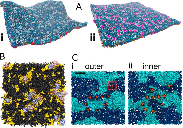
Protein-induced lateral heterogeneity. (A) Lateral heterogeneity induced by membrane proteins. The gp130 TMD (i), shown in red, gets solvated by GM3 (light blue) and PIP2 (orange). The S1P1 (ii), shown in purple, interacts favorably with PIP2 and cholesterol (green). Adapted with permission from ref (56). Copyright 2015 American Chemical Society. (B) Preferential solvation of dopamine D2 (blue) and adenosine A2A (red) receptors by SDPC (yellow) with a polyunsaturated chain. The other four lipid types are shown in gray. Reproduced with permission from ref (57). Copyright 2016 Guixà-González et al. (https://creativecommons.org/licenses/by/4.0/legalcode). (C) The formation of a Lo domain surrounded by the influenza hemagglutinin proteins due to sterical hindrance. DLiPC is shown in dark blue, DPPC in cyan. Adapted with permission from ref (605). Copyright 2013 Parton et al. (https://creativecommons.org/licenses/by/4.0/legalcode).

