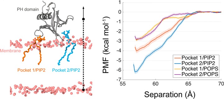
Figure 60.
PH domain binding to PIP2 lipids. (left) Simulation snapshot of a PH domain in the bound state with PIP2. The PH domain is shown in gray; DOPC phosphorus atoms are depicted as pink spheres. (right) Potential of mean force profiles for the PH domain of the ACAP1BAR-PH protein dimer bound to PIP2 at pocket 1 and pocket 2. Reproduced with permission from ref (1184). Copyright 2017 American Chemical Society.

