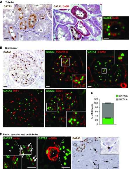
Figure 1.
Expression of GATA3 in healthy adult kidneys. (A) IHC for GATA3 (brown) in time-zero renal allograft biopsy samples from healthy adults demonstrated strong nuclear expression of GATA3 in a subset of tubular epithelial cells. CaSR (red) was used as a nephron segment marker53 in double staining, which showed that GATA3 was absent from proximal tubules (PT) and thick ascending limbs (TAL), but was expressed in distal/connecting nephron segments (DT/CN) and in principal cells of the CDs where it was absent from intercalated cells (arrow). IF staining for GATA3 (green) and CaSR (red) confirmed the cell specific expression of GATA3 in principal cells of the CD, which do not express CaSR. Scale bars, 20 μm. (B) Glomerular GATA3 expression was detected by IHC (brown) and by IF (green) where it was confined to MCs costaining with MC markers PDGFR-β and α-SMA (red). GATA3 did no colocalize with WT-1 or CD31, markers of epithelial (podocyte and parietal) and endothelial cells, respectively. Scale bars, 20 μm. (C) Quantification of the number of GATA3-expressing cells in GCS areas (n≥10) from healthy human kidneys (n=6). (D) GATA3 colocalized with the aspartyl-protease renin (red) in juxtaglomerular cells (arrows) and with α-SMA (red) in smooth muscle cells of the afferent arterioles (A). Peritubular expression of GATA3 was detected by IHC in a subset of interstitial fibroblasts (arrows). Arterioles indicated by dashed lines. Scale bars, 20 μm. Daltonized versions of Immunofluorescence pictures are provided in Supplemental Appendix 1. G, glomerulus.

