Figure 4.
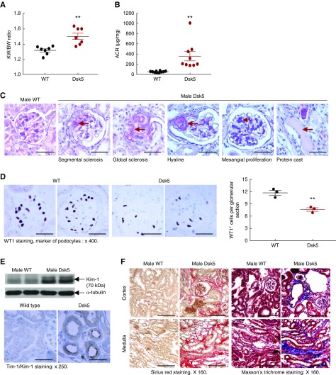
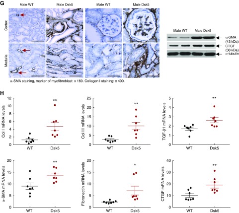
Male Dsk5 mice developed spontaneous kidney injury. Male wild-type and Dsk5 mice were euthanized at 15 weeks of age. (A) Male Dsk5 mice had kidney hypertrophy as indicated by increased kidney weight versus body weight (KW/BW) ratios (**P<0.01; n=7). (B) Male Dsk5 mice had increased albuminuria as indicated by increased urinary albumin-to-creatinine ratio (ACR) at 15 weeks of age (**P<0.01; n=10 in wild-type group and n=9 in Dsk5 group). (C) Period acid–Schiff staining showed that kidneys of male Dsk5 mice exhibited segmental and global glomerular sclerosis, mesangial expansion, hyaline, and protein casts. Original magnification, ×400 (scale bar, 25 µm). (D) Male Dsk5 mice had loss of podocytes as indicated by two representative photomicrographs from wild-type and Dsk5 mice (left panel) and quantitatively decreased WT1-positive cells (right panel; **P<0.01; n=3). Original magnification, ×400 (scale bar, 25 µm). (E) Male Dsk5 mice had epithelial cell injury as indicated by increased renal expression levels of KIM-1, a marker of proximal tubule epithelial injury. Original magnification, ×250 (scale bar, 40 µm). (F) Male Dsk5 mice exhibited tubulointerstitial fibrosis as indicated by Sirius Red and Masson trichrome staining. Original magnification, ×160 (scale bar, 62.5 µm). (G) Immunostaining and immunoblotting determined that male Dsk5 mice had increased renal expression levels of α-SMA, a marker of myofibroblasts, CTGF, and collagen I. Original magnification, ×160 for α-SMA (scale bar, 62.5 µm) and ×400 for collagen I (scale bar, 25 µm). Arrows indicate blood vessels. (H) Kidneys of male Dsk5 mice had increased mRNA levels of profibrotic and fibrotic components, including α-SMA, CTGF, TGF-β, collagen I and III (Col I and Col III), and fibronectin. *P<0.05; **P<0.01. n=7 in each group.


