Figure 6.
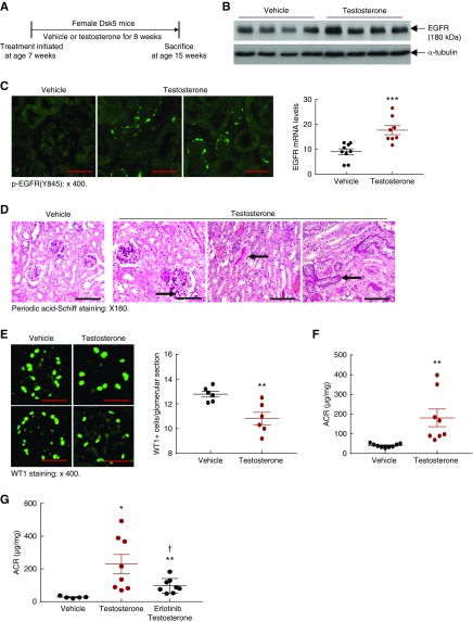
Testosterone supplementation led to increases in renal EGFR expression and injury in female Dsk5 mice. (A) Seven-week-old female Dsk5 mice were given testosterone at a dose of 10 mg/kg per day for 8 weeks and were euthanized at 15 weeks of age. (B and C) Testosterone administration led to increases in renal EGFR activation, mRNA, and protein levels (***P<0.001 versus vehicle; n=9 in vehicle group and n=8 in testosterone group). Original magnification, ×400 (scale bar, 25 µm). (D) Testosterone treatment led to mesangial expansion, protein casts, and tubular epithelial cell atrophy in female Dsk5 mice (arrows). Original magnification, ×160 (scale bar, 50 µm). (E) Testosterone administration caused loss WT1 staining, a marker of podocytes, in female Dsk5 mice (**P<0.01; n=6). Original magnification, ×400 (scale bar, 25 µm). (F) Testosterone supplementation led to increased albuminuria in female Dsk5 mice (**P<0.01; n=9 in vehicle group and n=8 in testosterone group). (G) Testosterone-induced albuminuria in female Dsk5 mice was prevented by erlotinib, an inhibitor of EGFR tyrosine kinase activity (*P<0.05, **P<0.01 versus vehicle group; †P<0.05 versus testosterone group; n=5 in vehicle group and n=8 in testosterone and testosterone plus erlotinib groups).

