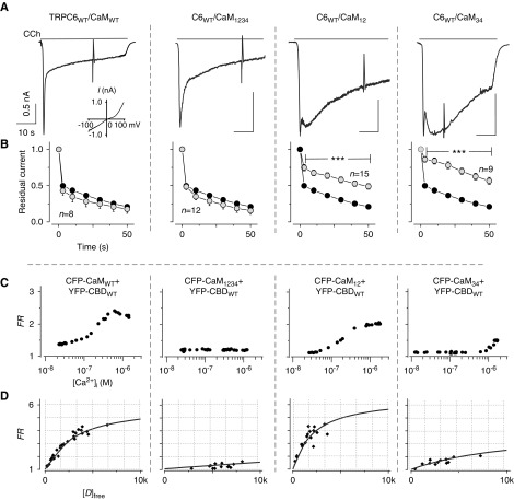
Figure 1.
Functional analysis of CaM mutants and Ca2+-dependent binding. (A) Representative currents recorded from HEK293 cells coexpressing with TRPC6 and CaMWT or a CaM mutant (CaM12, CaM34, or CaM1234). Muscarinic receptor (M1R) is also coexpressed for the electrophysiologic experiments in HEK293 cells. The inset shows typical I–V curve of TRPC6. (B) Fractions of residual currents plotted as a function of time after the peak. Black circles indicate control data from TRPC6 with M1R-transfected cells only (n=14, CaMendo), here and throughout. (C) Representative profiles of Ca2+-dependent binding of CaMs to the TRPC6 CBD. FRET measurement is explained in the Methods section. (D) Quantification of the affinity of binding of CBDWT to CaM mutants by plotting donor free ([D]free) concentrations against FRET signal (FR). Data from various cells were collected at their maximum FR values in (C). Lines represent fitting of CBDWT versus CaMMUT. FRET data are summarized in Supplemental Table 1.

