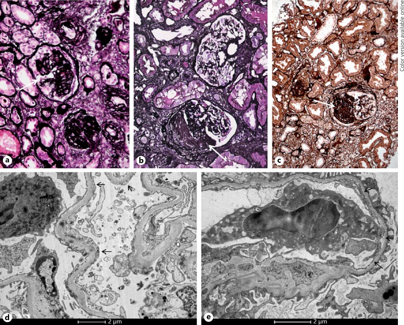
Fig. 1.
Kidney biopsy images in the 3 cases. Light microscopy (Jones staining) showed glomeruli with segmental sclerosis (arrows) in case 1 (a), case 2 (b), and case 3 (c). Electron microscopy of case 1 showed partial foot process effacement, with areas of intact foot processes (*) alternating with areas with foot process effacement (arrows, d). In addition to partial foot process effacement, EM of case 2 also showed a thin GBM thickness with a mean of 252 nm (arrows, e).

