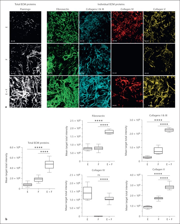
Fig. 1.
Co-culture of epithelial cells and fibroblasts leads to spontaneous ECM production. Epithelial cells and fibroblasts were cultured for 7 days in mono- or co-culture (1:1 ratio). Cells were then lysed; ECM was fixed and stained using a total protein dye or specific antibodies. a Images of total ECM components (total ECM proteins, white;), and individual ECM components (fibronectin, green; collagens I and III, blue; collagen IV, red; collagen V) are representative of 4 independent experiments. b Graphs show the results obtained after the quantification of the fluorescent signal from the images. The box and whisker plots show one representative out of 4 independent experiments with 12 replicate wells per condition. E, mono-culture of epithelial cells; F, mono-culture of fibroblasts; E + F, co-culture of epithelial cells and fibroblasts. Images show a single field. Scale bars represent 100 µm. * p < 0.05, ** p < 0.01, *** p < 0.001, **** p < 0.0001, one-way ANOVA vs. co-culture. ns, not significant; ECM, extracellular matrix.

