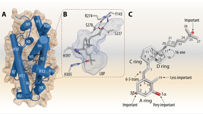
Figure 2.
1,25(OH)2D3 complexed to the VDR-LBD. The VDR-LBD has a conserved 3D architecture, which is made of a three-layer α-helical sandwich. In the lower part of the LBD the LBP is located. All the helices are labeled from N-terminus toward C-terminus and numbered in white color (A). Details on the LBP with bound 1,25(OH)2D3 and critical amino acids that provide anchoring contacts for the three OH groups (B). Details on the conformation of the bound 1,25(OH)2D3 molecule with the annotated OH groups and highlights to its contribution of its activity. The numbering of the carbons atoms is indicated (C). The figure is based on the PDB code 1DB1.

