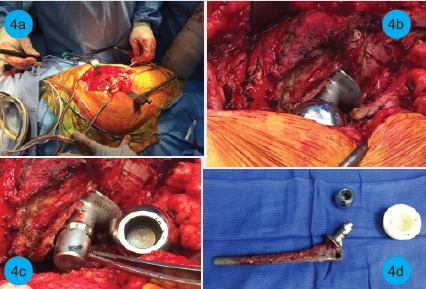
Figure 4.

Intraoperative images demonstrate metallosis and evidence of trunnionosis. The approach to the implant (a), operative view of head and tissue around it (b), inspection of the neck showing trunnion wear with a black discoloration at the tip of the taper (c), and explanted femoral implant, head, and liner (d) are all shown.
