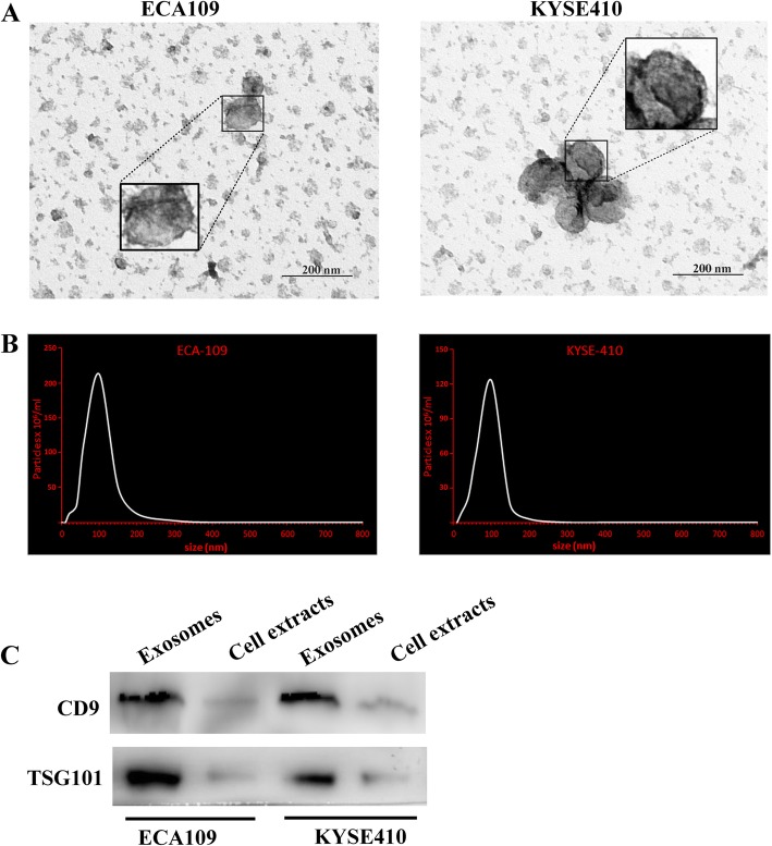
Fig. 1.
Identification of the purified extracellular vesicles. a Transmission electron micrographs of extracellular vesicles derived from ECA109 and KYSE410. b The nanoparticle concentration and size distribution of the extracellular vesicles derived from ECA109 and KYSE410. c The expression level of CD9 and TSG101 (exosome specific markers) in extracellular vesicles

