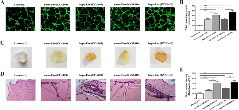
Fig. 4.
Hypoxic exosomes promoted angiogenesis in vitro and increased the vessel density in vivo. HUVECs were plated with matrigel and cultured with exosomes (25 μg /mL) or not. Representative pictures of tube formation were taken after stained with Calcein-AM (a). The tube formation ability was quantified by measuring the total branching length (b). Matrigel containing exosomes, or not, were injected subcutaneously into the nude mice. Representative images of the general observation of matrigel plugs (c). In vivo neovascularization induced by exosomes was measured by H&E staining. Representative pictures of neovascularization were shown in (d) and quantified for blood vessel density (e). Data was presented as mean ± standard deviation (SD). *P < 0.05, **P < 0.01, ***P < 0.001

