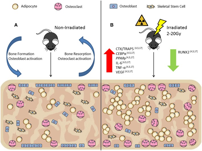
Figure 2.
In vivo rodent models show irradiation alters the bone marrow microenvironment by increasing osteoclast numbers per bone surface (Oc.S/BS) and decreasing osteoblast numbers per bone surface (Ob.S/BS) resulting in decreased trabecular bone volume with a rapid influx of bone marrow adipocytes. (A) Non-irradiated control, demonstrates normal bone turnover processes. (B) Irradiated (2-20Gy), demonstrates the uncoupling of the bone formation/resorption ratio through increased CTX/TRAP5 (osteoclast activity) and decreased RUNX2 (osteoblast activity) expression. In vivo irradiation exposure also has increased CEBPα and PPARγ (adipogenesis markers), and IL-6, TNF-α, and VEGF (inflammatory and senescent markers).

