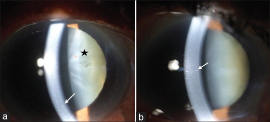
Figure 1.

(a) Anterior segment photograph of the right eye showing medium-sized keratic precipitates on the endothelium (white arrow). Pigments can be seen over the anterior capsule of the lens (star). (b) Anterior segment photograph of the left eye showing medium-sized keratic precipitates on the endothelium (white arrow)
