Goltz syndrome is a rare multisystem disorder affecting tissues of meso-ectodermal origin with cutaneous, skeletal, ocular, and dental abnormalities.
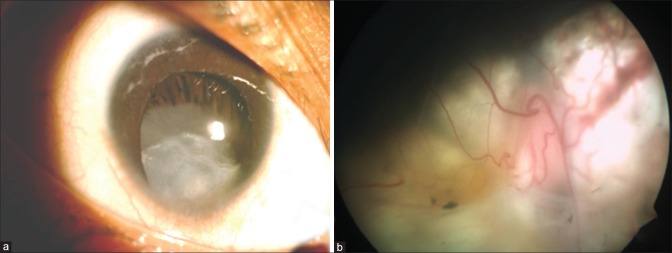
A 3-year-old girl presented with decreased vision in both the eyes and a history of cleft-lip repair and delayed milestones. Her vision was 3/60 by Lea symbols in both eyes. There was a partial ptosis of right eyelid, microcornea, microphthalmos, and iris coloboma in both the eyes with subluxated cataractous lens (right more than left) and stretched ciliary processes [Fig. 1]. The fundus examination revealed retinochoroidal coloboma involving disc and macula in both the eyes.
Figure 1.
(a) Ophthalmic examination showed microcornea, microphthalmos with coloboma iris, and subluxated cataractous lens with stretched ciliary processes. (b) Retinochoroidal coloboma seen in the fundus photograph of right eye
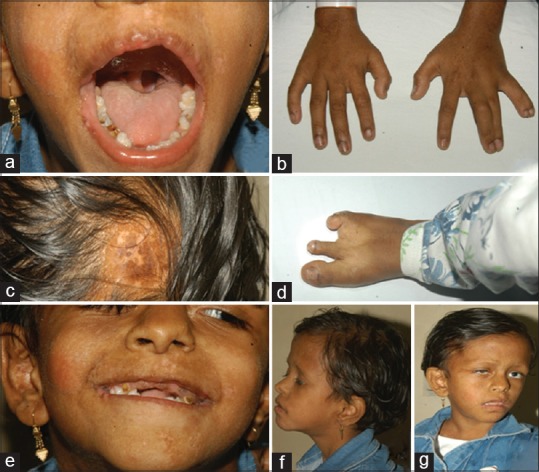
There was microcephaly and alopecia with asymmetry of both sides of the face along with notched left nares. She had hypopigmented, depressed, and ill-defined macules dispersed over her face. There was polydactyly with syndactyly of the fifth and sixth finger, and the third and fourth finger of left hand, with a characteristic lobster claw deformity; oligodactyly of right third and fourth toe; and hypoplasia of the nails [Fig. 2]. Her X-ray spine showed an abnormal sacral vertebra. A high-arched palate, small oropharyngeal papilloma, abnormal dentition with enamel defects, abnormal spacing, malocclusion, and gingival hyperplasia were also noted. Heterozygous nonsense mutation c.727C>T (p.R243*) in exon 9 in the DNA of baby was found and has been earlier described in patients with Goltz-Gorlin syndrome.
Figure 2.

Composite picture depicting various manifestations of a child with Goltz syndrome. (a) External picture of face showing repaired cleft lip (yellow arrow), high-arched palate, small oropharyngeal papilloma, abnormal dentition with abnormal spacing, malocclusion, gingival hyperplasia, and enamel defects (black arrow). Hypopigmented macules over her face (white arrow) and body. (b) Polydactyly, syndactyly (white arrows) of left-hand fingers with lobster-claw deformity. (c) Alopecia. (d) Oligodactyly of right toes (arrow) with hypoplasia of the nails. (e-g) Asymmetry of two sides of face with notched nare on left side and microcephaly
Goltz syndrome is an uncommon X-linked disorder with a probable locus at PORCN gene (Xp11.23). Skin involvement is essential for the diagnosis with hypoplasia of the dermis with ocular involvement seen in 40–70% patients.[1,2,3,4] Some patients present with mental retardation, microcephaly, and hearing defects. Skeletal defects are seen in 80% of the patients, including spinal defects, polydactyly, syndactyly, or clinodactyly.[5]
All features may not be present in one patient due to mosaicism. Treatment of Goltz syndrome is largely supportive. Ophthalmologist may be the first point of contact; timely visual rehabilitation may reduce the associated morbidity of the disease.
Declaration of patient consent
The authors certify that they have obtained all appropriate patient consent forms. In the form the patient(s) has/have given his/her/their consent for his/her/their images and other clinical information to be reported in the journal. The patients understand that their names and initials will not be published and due efforts will be made to conceal their identity, but anonymity cannot be guaranteed.
Financial support and sponsorship
Nil.
Conflicts of interest
There are no conflicts of interest.
References
- 1.Goltz RW, Peterson WC, Gorlin RJ, Ravits HG. Focal dermal hypoplasia. Arch Dermatol. 1962;86:708–17. doi: 10.1001/archderm.1962.01590120006002. [DOI] [PubMed] [Google Scholar]
- 2.Goltz RW. Focal dermal hypoplasia syndrome: An update. Arch Derm. 1992;128:1108–11. [PubMed] [Google Scholar]
- 3.Thomas JV, Yoshizumi MO, Beyer CK, Craft JL, Albert DM. Ocular manifestations of focal dermal hypoplasia syndrome. Arch Ophthalmol. 1977;95:1997–2001. doi: 10.1001/archopht.1977.04450110091009. [DOI] [PubMed] [Google Scholar]
- 4.Gisseman JD, Herce HH. Ophthalmologic manifestations of focal dermal hypoplasia (Goltz syndrome): A case series of 18 patients. Am J Med Genet Part C Semin Med Genet. 2016;172C:59–63. doi: 10.1002/ajmg.c.31480. [DOI] [PubMed] [Google Scholar]
- 5.Knockaert D, Dequeker J. Osteopathiastriata and focal dermal hypoplasia. Skelet Radiol. 1979;4:223–7. doi: 10.1007/BF00347218. [DOI] [PubMed] [Google Scholar]


