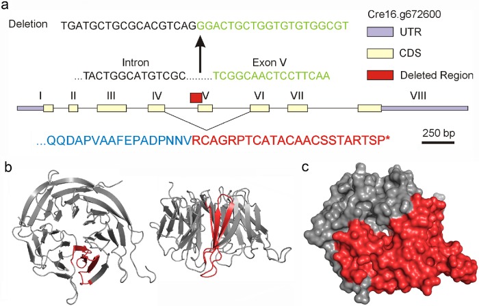
FIGURE 3:
Analysis of the wdr92-1 insertional mutant. (a) Map of the WDR92 intron/exon gene structure showing the region (red box) and sequence deleted by insertion of the C1B1 paromomycin resistance cassette. Removal of the 5′ splice site for exon V allows exon IV to splice to exon VI, leading to a frameshift, 23 new residues, and a stop codon (red in lower sequence). (b) Two views of the ribbon structure (PDB 3I2N) for human WDR92 illustrating in red the blade of the β-propeller that is encoded by exon V. (c) Surface rendering of the WDR92 structure indicating in red the C-terminal region missing in the encoded wdr92-1 mutant.

