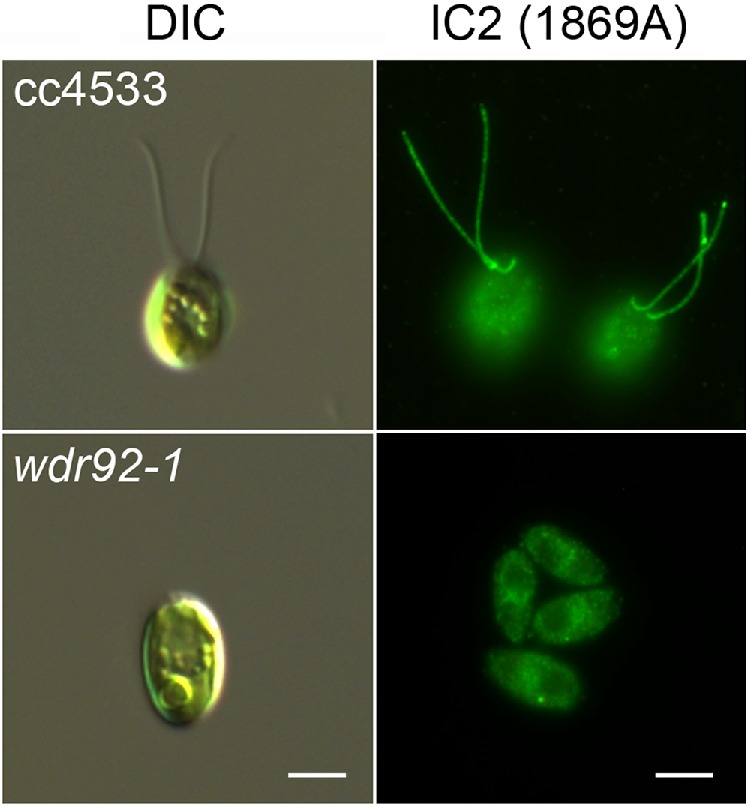
FIGURE 4:

wdr92-1 mutant cells lack cilia. Left, differential interference contrast (DIC) images of control (cc4533) and wdr92-1 cells. The mutant lacks obvious ciliary structures. Right, immunofluorescence micrographs of cc4533 and wdr92-1 cells probed with monoclonal antibody 1869A that specifically recognizes outer arm dynein IC2 (King et al., 1985). No obvious staining of ciliary structures or stubs was observed. Scale bars: 5 μm.
