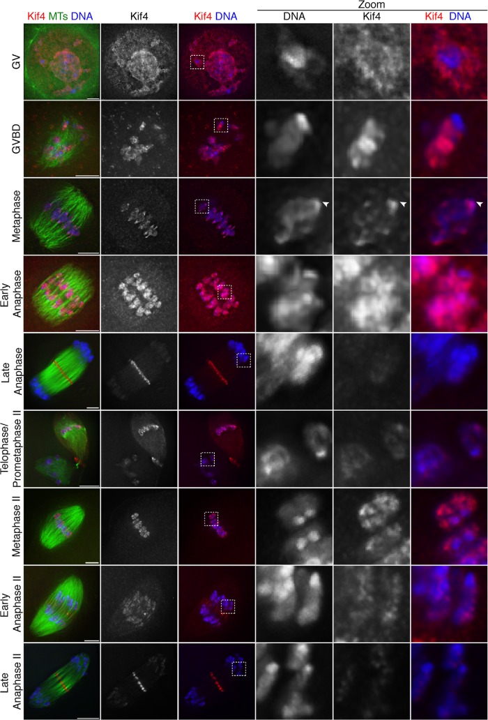
FIGURE 1:
Dynamic Kif4 localization in mouse oocytes. Representative deconvolved images of the major stages of mouse oocyte meiosis I and II. Shown are Kif4 (red), microtubules (green), and DNA (blue). Kif4 is chromosome-associated through early anaphase and then largely leaves the chromosomes, which is highlighted by the zoomed regions. Early in prometaphase, Kif4 appears excluded from the region near the ends of bivalents, where kinetochores are located, but spreads to this region by metaphase (arrowhead). All images are partial projections chosen to highlight chromosomal staining. Bars = 5 µm.

