FIGURE 2:
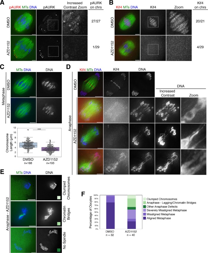
Aurora B/C regulate Kif4’s localization and inhibition of these kinases causes a variety of meiotic defects. (A) Metaphase I spindles stained for phospho-Aurora kinase A/B/C (pAURK; red), microtubules (green), and DNA (blue) in DMSO control and 100 nM AZD1152-treated oocytes. Although pAURK pole staining (primarily Aurora kinase A) is unaffected, pAURK fluorescence on the chromosomes is reduced. Although the chromosomal staining appears dim in the middle column, given the brighter pole staining, this difference is more apparent in the zoom of the chromosomal region where we increased contrast (right column). (B) Metaphase I spindles stained for Kif4 (red), microtubules (green), and DNA (blue) in DMSO control and 100 nM AZD1152-treated oocytes. Kif4 fluorescence is reduced following AZD1152 treatment, highlighted in the zoom. For A and B, numbers at the right indicate the number of images examined that showed substantial pAURK or Kif4 chromosomal staining. (C) Metaphase spindles stained for microtubules (green) and DNA (blue) showing change in chromosome alignment and bivalent structure following AZD1152 treatment. Quantification of chromosome lengths is shown below the images. Average chromosome length is 5.2 µm ± 0.1 (n = 188 chromosomes from 11 DMSO-treated spindles) and 3.5 µm ± 0.1 (n = 193 chromosomes from 10 AZD1152-treated spindles), demonstrating that chromosomes are shorter following AZD1152 treatment. Asterisks denote p < 0.05. (D) Anaphase I spindles stained for Kif4 (red), microtubules (green), and DNA (blue). AZD1152 treatment causes mislocalization of Kif4 in anaphase and chromosome segregation errors with lagging chromosomes and chromatin bridges; images in the right two columns have increased contrast to highlight these features. (E) Examples of other anaphase defects, stained for microtubules (green) and DNA (blue), following AZD1152 treatment, including clumped chromosomes (top row) and chromatin bridges and/or aberrant spindles (bottom two rows) color-coded to reflect the graph in F. (F) Quantification of spindles from two independent experiments; oocytes were matured for 6 h, at which time most control oocytes are in metaphase with mild to no chromosome misalignment. AZD1152-treated oocytes show increased chromosome congression defects (categories displayed in light purple; misaligned metaphase with one to two chromosomes misaligned or severely misaligned with more than three chromosomes misaligned), enter anaphase prematurely (categories displayed in green), and show a variety of anaphase defects with examples shown in E. Bars = 5 µm.

