FIGURE 6:
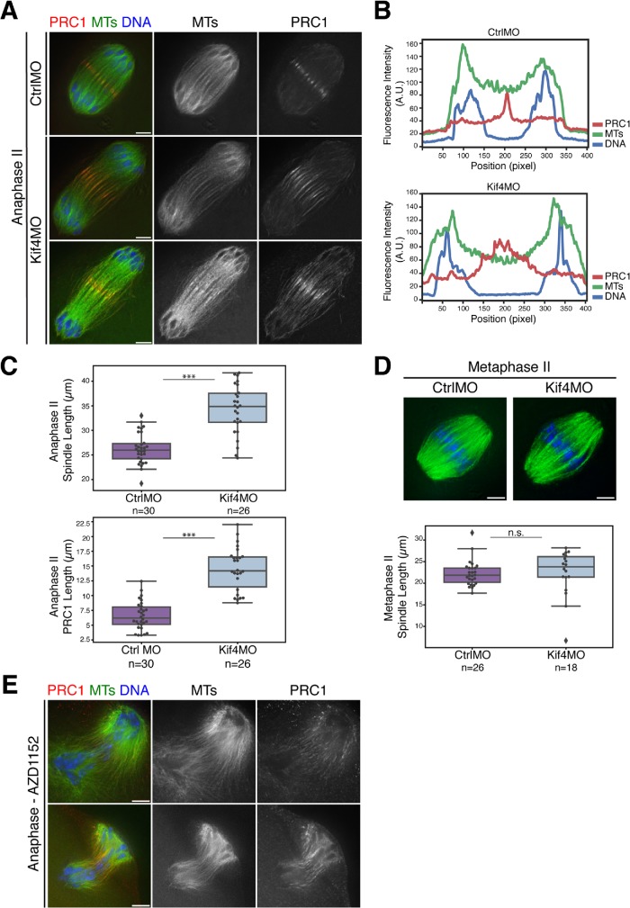
Anaphase spindle length and midzone organization is regulated by Kif4 in oocytes. (A) Anaphase II spindles from oocytes injected with either control or Kif4 morpholinos stained for PRC1 (red), microtubules (green), and DNA (blue). Following Kif4 depletion, PRC1 does not form distinct structures and instead is spread on an expanded region of the anaphase spindle. (B) Line scans of the top two images from panel A, showing a tight peak of PRC1 in the control compared with a broader peak following Kif4 depletion. (C) Quantification of anaphase II spindle lengths (top) and PRC1 midzone spread (bottom); spindles are longer following Kif4 depletion and PRC1 localizes to a broader domain. Scatter plot overlaid on box plot shows individual spindle or PRC1 length measurements. Average anaphase II spindle lengths were 26.2 µm ± 0.6 (n = 30) for control-injected oocytes and 34.2 µm ± 1.0 (n = 26) for Kif4MO-injected oocytes. Average length of the PRC1 midzone is 6.6 µm ± 0.4 (n = 30) for the control and 14.5 µm ± 0.7 (n = 26) for Kif4MO. Conditions are significantly different (p < 0.0005, denoted by three asterisks). (D) Metaphase II spindles stained for microtubules (green) and DNA (blue). No major congression errors or spindle abnormalities were observed in 26 control and 18 Kif4-depleted spindles. Quantification of metaphase II spindle lengths are below the images; Kif4 depletion does not significantly affect metaphase II spindle length. As in C, box plots with overlaid scatter plots show individual spindle length measurements. No significant difference (p > 0.05, denoted by n.s.) between the two conditions. (E) AZD1152-treated anaphase I spindles stained for PRC1 (red), microtubules (green), and DNA (blue). In 10/10 aberrant anaphase spindles, there were disorganized midzones and mislocalized PRC1 with AURKB/C inhibition (two representative images shown). Bars = 5 µm.

