FIGURE 1:

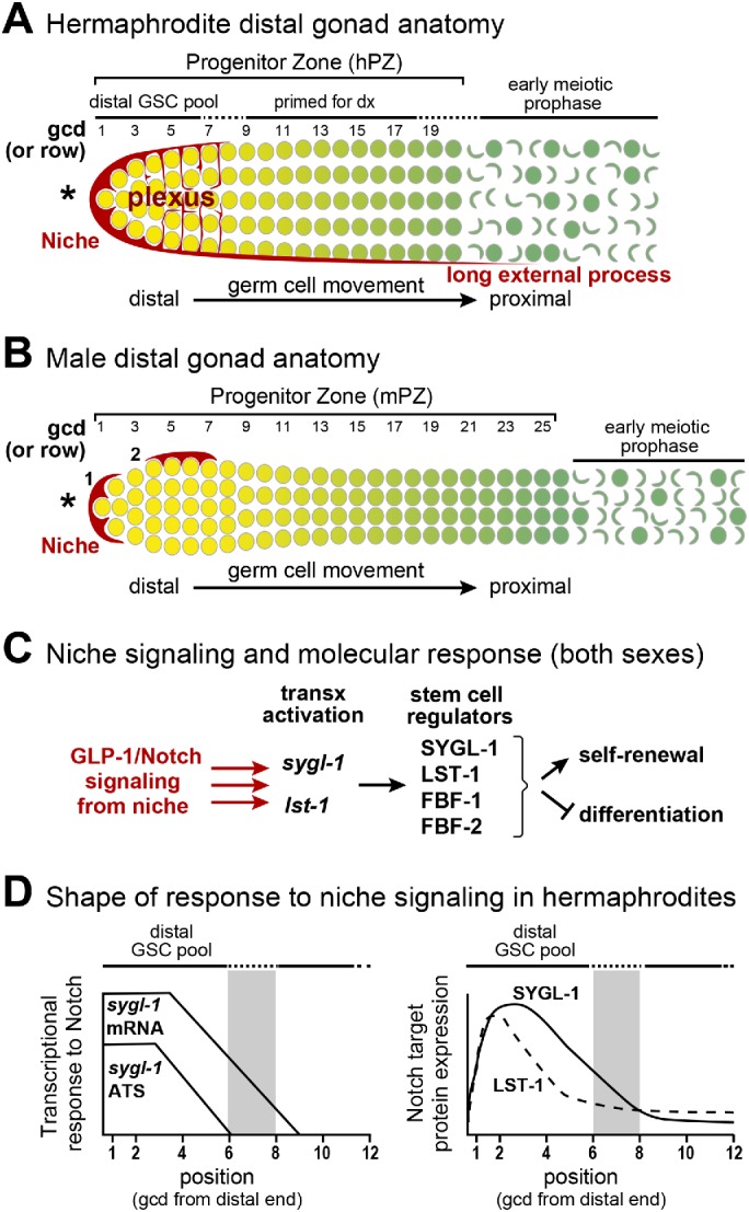
Hermaphrodite and male DTC niches and PZs (PZs). (A, B) Diagrams of the distal gonads of each sex, each with a mesenchymal niche (red) and a germline PZ with ∼225 germ cells that develop as they move from distal to proximal along the gonadal axis (yellow to green). Germ cell position is conventionally described as the number of germ cell diameters (gcd) from the distal end, with rows of germ cells at each position. Germ cells are depicted at the gonad periphery; not shown is the central rachis of cytoplasm to which all germ cells connect via bridges (Hirsh et al., 1976; Amini et al., 2014; Seidel et al., 2018). Although germ cells are connected through the rachis, each is partially enclosed by plasma membrane and functions largely autonomously with respect to cell cycle stage; for simplicity, the convention is to call them germ cells. Also not shown are germline folds in the hermaphrodite PZ (Raiders et al., 2018; Seidel et al., 2018). Here and in all figures, an asterisk marks the distal end. (A) Hermaphrodite distal gonad. The hDTC cell body caps distal germ cells and extends a plexus of processes that contact most germ cells through rows 6–8 (Byrd et al., 2014; Lee et al., 2016). The hPZ has fewer cells per row distally and then widens (Morgan et al., 2010); it extends ∼20 gcd from the distal end. The hPZ contains a distal pool of naïve, stem cell–like undifferentiated cells (yellow), and a proximal pool of cells that are primed for differentiation and mature progressively (graded green) as they move proximally (darker green) (Cinquin, Crittenden, et al., 2010; Fox et al., 2011). (B) Male distal gonad. One mDTC typically resides at the distal end and the other on the dorsal side. We refer to the dorsal mDTC as lateral since in extruded gonads its dorsal, lateral, or ventral position cannot be determined. The mPZ bulges distally and then becomes thinner, extending ∼25 gcd from the distal end (Morgan et al., 2010). Other features shown for the hermaphrodite distal gonad (e.g., distal and proximal pools) were not known prior this work. Here and in all figures, the position of most-distal male DTC is marked 1 and the lateral DTC is marked 2. (C) Molecular stem cell regulation. The niche uses GLP-1/Notch signaling to activate transcription of the sygl-1 and lst-1 genes in GSCs (Kershner et al., 2014; Lee et al., 2016). The SYGL-1 and LST-1 proteins work with FBF-1 and FBF-2 to promote self-renewal and inhibit differentiation (Shin, Haupt, et al., 2017). (D) Hermaphrodite GSC response to GLP-1/Notch signaling, based on previous reports (Lee et al., 2016; Shin, Haupt, et al., 2017). Left, sygl-1 ATS reveal the direct response to signaling; sygl-1 mRNAs extend more proximally than ATS. Both ATS and mRNAs are restricted to the GSC pool region and are graded. Right, SYGL-1 (solid line) and LST-1 (dashed line) proteins are restricted to the GSC pool region and graded. Gray bar indicates the estimated proximal boundary of the GSC pool (6–8 gcd; Cinquin, Crittenden, et al., 2010).
