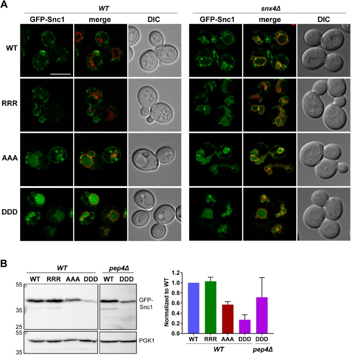
FIGURE 4:
Positively charged residues within the Snc1 transmembrane domain prevent targeting to the vacuole lumen. (A) Micrographs of wild-type or snx4Δ cells expressing wild-type or indicated GFP-Snc1 mutants. GFP-SNC1 genes were integrated into the URA3 locus to prevent plasmid expression variability. Vacuoles are visualized using FM4-64 dye. Scale bar indicates 5 μm. (Β) Cell lysates of wild-type or pep4Δ cells expressing the indicated GFP-Snc1 variants were probed with anti-GFP antibody and the amount of GFP-Snc1 was determined by semiquantitative Western blot analysis. Anti-PGK1 immunoblot was used as protein loading control. The positions of molecular mass (kDa) markers are indicated. The results from three experiments were averaged and standard error of the mean indicated.

