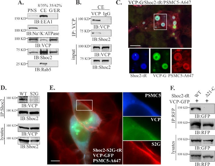
FIGURE 2:
VCP is in complex with Shoc2 on endosomes. (A) Post nuclear supernatants (PNSs) from Cos1 cells were layered on 8–42% sucrose gradients and subjected to ultracentrifugation. The indicated proteins were identified by immunoblotting (IB) using specific antibodies in crude endosome (CE) or Golgi and endoplasmic reticulum (G/ER) fractions. (B) VCP was immunoprecipitated from the crude endosomal fraction and analyzed by using specific anti-VCP and Shoc2 antibodies. (C) Cos-SR cells depleted of endogenous Shoc2 and stably expressing Shoc2-tRFP were transfected with VCP-GFP and GST-PSMC5, permeabilzed with 0.05% saponin and then fixed, immunostained for GST, and followed by immunofluorescence microscopy. Insets show high-magnification images of the region indicated by white rectangles. Scale bars: 10 μm. (D) Shoc2 was immunoprecipitated from Cos-SR cells depleted of endogenous Shoc2 and stably expressing WT or S2G mutant Shoc2-tRFP. The immunoprecipitates were analyzed with anti-VCP and -Shoc2 antibodies. (E) Cos1 cells stably expressing the Shoc2-S2G mutant (Shoc2-S2G-tR) were transfected with GST-PSMC5 and VCP-GFP. GST-PSMC5 was immunostained with anti-GST antibody. Cells were fixed and followed by immunofluorescence microscopy. Insets show high-magnification images of the regions of the cell indicated by white rectangles. Scale bars: 10 μm. (F) Cos1 cells depleted of endogenous Shoc2 and stably expressing either full-length Shoc2-tRFP (WT) or the mutant of Shoc2 (Δ21-C) were transfected with VCP-GFP. Shoc2 was immunoprecipitated using anti-RFP antibodies. The immunoprecipitates were analyzed by immunoblotting using anti-RFP and -GFP antibodies. The results in each panel are representative of those from three independent experiments.

