Figure 6.
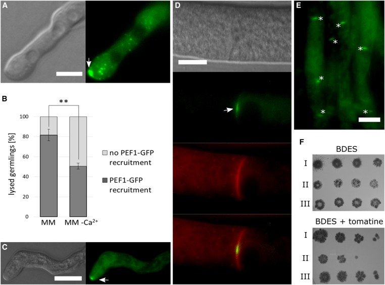
PEF1-GFP is recruited in response to tomatine-induced cell lyses and contributes to the survival in presence of this pore-forming drug. (A) Recruitment of PEF1-GFP to the germling tips in response to tomatine-induced cell lysis (arrow). Left: DIC; right: GFP fluorescence. Bar, 5 µm. (B) Quantification of PEF1-GFP recruitment in germlings treated with tomatine on MM and MM without Ca2+. Error bars indicate the SD calculated from three independent experiments (n = 100 each). The asterisks represent statistically significant differences determined by the Student’s t-test (**P ≤ 0.01). (C) Recruitment of PEF1-GFP to a tip of a mature hypha in response to tomatine-induced cell lysis (arrow). Left: DIC; right: GFP fluorescence. (D) PEF1-GFP recruitment to the septal pore (arrow) after treatment with tomatine in mature hypha. The plasma membrane was stained in red with the lipophilic dye FM4-64. Top image: DIC; second image from top: GFP; third image from the top: FM4-64; bottom image: merger of GFP and FM4-64 images. (E) Overview of a group of hyphae exhibiting PEF1-GFP at septal pores after tomatine treatment (aggregates indicated by asterisks). (A–E) Strain GN9-22 (Ptef-1-pef-1-gfp). Bars in (A–E), 10 µm. (F) Fivefold serial spore dilutions (105–102) of wild type (I) (FGSC 988), ∆pef1 (II) (FGSC 15890), and the complemented strain ∆pef1 Ptef-1-pef1-gfp (III) (GN9-22) were spotted on BDES medium and on BDES medium containing 75 µg/ml tomatine. Growth was documented after 3 dpi.

