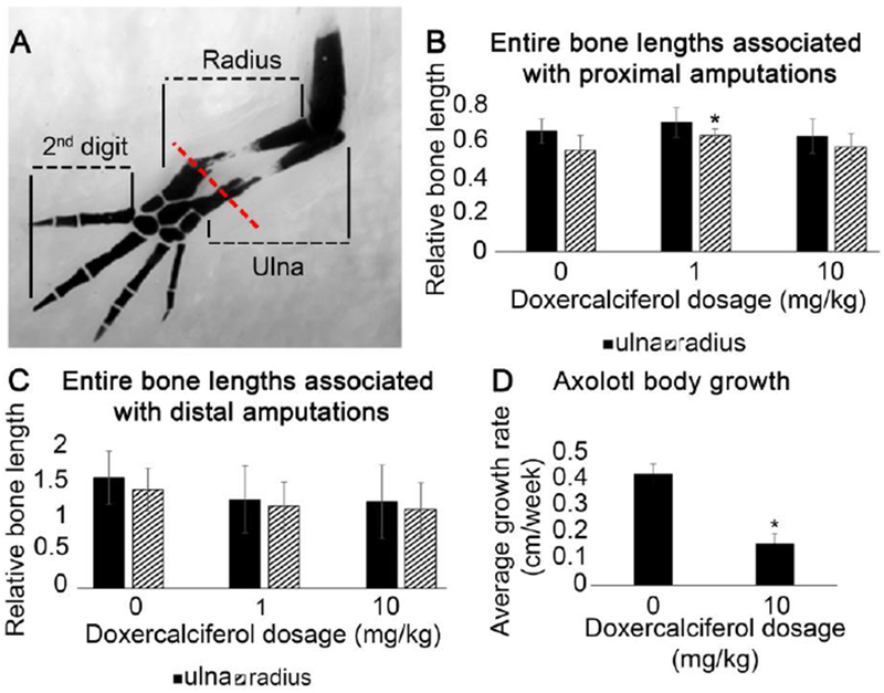
Figure 3: Doxercalciferol had minimal effect on regenerating bone length.

(A) Representative image showing how the entire radius and ulna bone were quantified, along with the second digit, in DMSO and Doxercalciferol treated forelimb regenerates. Red line represents the site of amputation. Histogram representing the proportional length of the radius and ulna (measured as in “A”) resulting from (B) proximal amputations (N = 13 for DMSO treated amputations, N = 10 for 1mg/kg and 8 for 10mg/kg Doxercalciferol treatment groups; one-way Anova analysis p<0.05, post hoc Tukey tests revealed that DMSO treated radia were significantly shorter than 1mg/kg Doxercalciferal treated radia) or (C) distal amputations (N = 7 for DMSO treated amputations, N = 10 for both Doxercalciferol treatment groups, one-way Anova analysis p>0.05). Error bars represent standard deviation of the mean. (D) Body length (snout-to-tail tip) was measured at the day of injection and then 8 weeks later. The average change in body length over this time period was significantly smaller for Doxercalciferol treated axolotl (N=5) compared to vehicle treated animals (N=9) (t-test, p<0.05).
