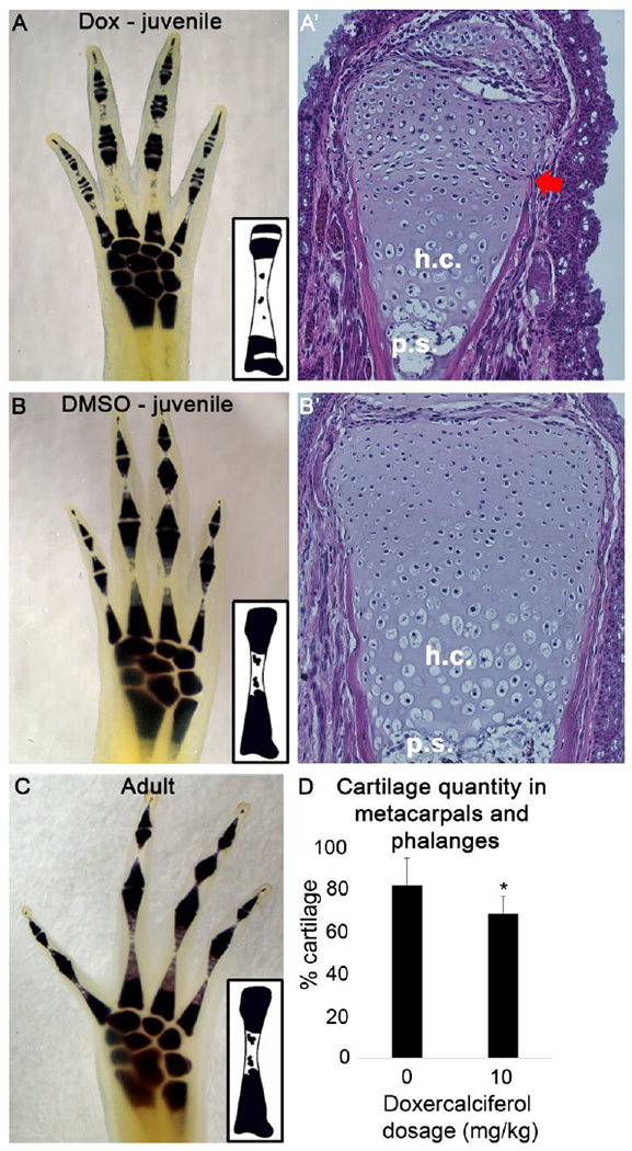
Figure 5: Doxercalciferol treatment altered epiphysis structure in uninjured limb tissue.

Whole mount cartilage staining revealed that Doxercalciferol decreased metacarpal and phalange cartilage staining in mature autopods (A) relative to age matched, DMSO-treated controls (B). This observation was observed in all analogue treated autopods (N = 9). Hematoxylin and Eosin staining of the distal joint of the metacarpal in Doxcercalciferol- (A’) and DMSO-treated (B’) animals revealed unusual morphology in the epiphysis with Doxicalciferol tretament (red arrow). h.c. – hypertrophic chondrocyte,.s. - primary spongiosa. (C) Older, untreated animals (1 year old, N = 4), had a similar cartilage staining pattern as DMSO treated, juvenile animals. Insets: graphical representations of cartilage staining in the metacarpals. (D) Histogram representing the % of the metacarplas and phalanges that stains positive for cartilage. Doxercalciferol treatment significantly decreased the cartilage staining of the metacarpals and phalanges of mature autopods (t-test, p<0.05, N = 9 for both groups).
