
Fig. 1.
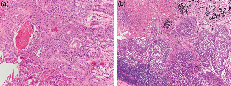
Pathological images. (a) Lung squamous cell carcinoma was identified by haematoxylin and eosin (HE) staining of primary tumour at the root of left lower lobe (magnification ×200). (b) Subcarina lymph nodes involvement was indicated by HE staining (magnification ×100).