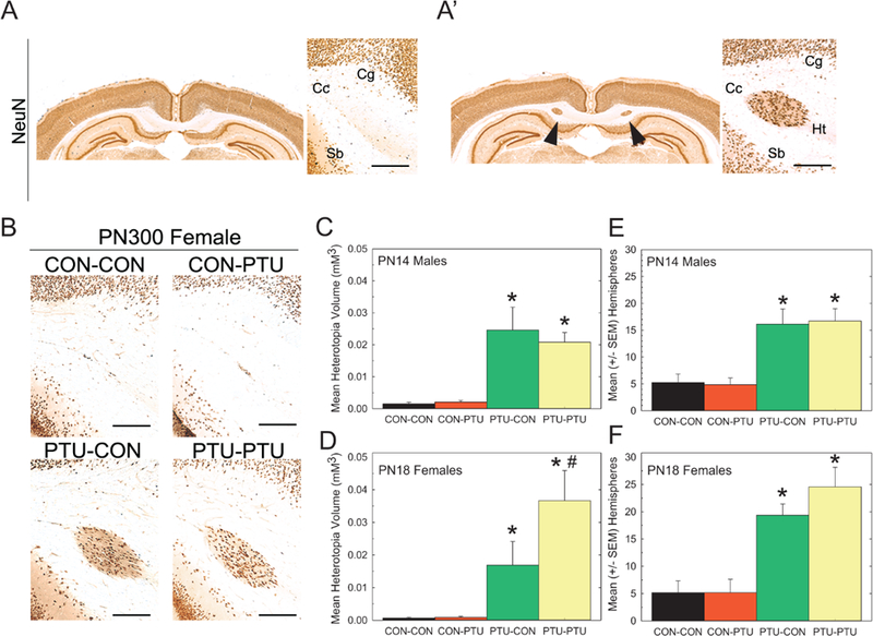
Figure 4.

(A) Coronal section of a CON-CON and (A’) PTU-PTU exposed pup showing presence and anatomical position of a heterotopia in NeuN-immunostained section. (B) Heterotopia were present in pups from PTU-CON and PTU-PTU treatment conditions, but not in controls or pups with exposure beginning on PN2 (CON-PTU). (C,D) Mean (±SE) heterotopia volume determined stereologically and the number of hemispheres a heterotopia was observed in male offspring on PN14 was higher in treatments with prenatal exposure (PTU-CON and PTU-PTU) than in control. (E,F) Similarly, heterotopia volume and hemisphere incidence was greater in PN18 females with prenatal exposure, suggesting that gestational exposure is necessary for heterotopia formation. Scale bar=50 mm. Cg, cingulum; Cc, corpus callosum; Sb, subiculum; Ht, heterotopia. Duncan’s t-test following significant ANOVA. *Significantly different from CON-CON; #significantly different from PTU-CON, p<.05.
