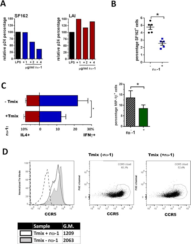
Fig 5. DCs exposed to rω-1 induce a Tmix cell population less susceptible to SF162 (R5) but not LAI (X4) infection.

(A) Percentage of SF162 or LAI infected cells in Tmix cell populations induced by rω-1 unexposed DCs was set to 100% and the level of infection found in Tmix cell cultures induced by rω-1 exposed DCs was expressed as a percentage of this 100%. (B) The percentage of SF162+ cells found in Tmix cell populations induced by unexposed DCs versus the percentage found Tmix cell populations induced by rω-1 exposed DCs (left panel) with +/- SE shown, with the same shown for LAI (right panel) with +/- SE depict. (C) Average percentage (± SEM) of IL-4, IFN-γ and MIP-1β producing T-cells in Tmix cell cultures induced by unexposed or rω-1 exposed DCs and depict with SD. (D) Percentage of cells with high CCR5 expression on their surface as well as the quantity of CCR5 per cell (geometric mean) for Tmix cell cultures induced by unexposed or rω-1 exposed DCs (dotted line unstained control), representative figure of 3 independent experiments. (B, C) Data from four independent experiments (± SD) using four different donor combinations where DCs were matured in the presence of 2–4μg/ml rω-1. *p<0.05.
