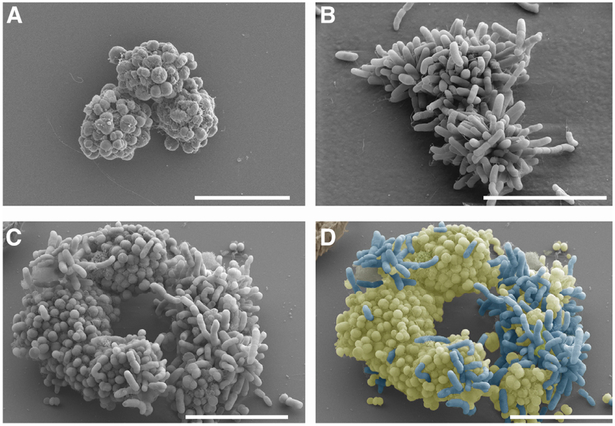
Fig 1. Nel and Ngo microcolonies associate with each other.
Scanning electron micrograph of (a) Ngo and (b) Nel cultured alone, and (c, d) Ngo and Nel cultured together for 5 h. The image in (c) was pseudocolored to discriminate Ngo (cocci; yellow) from Nel (rods; blue). Starting CFUs of each species: 5×10⁷. Scale bars: 10 μM.

