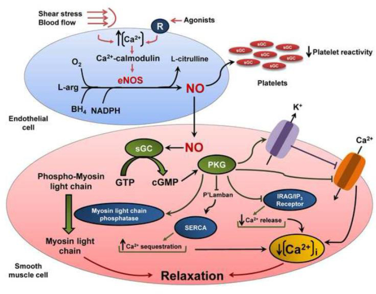
Figure 1.
Mechanisms involved in NO formation and vasodilation. Increases in intracellular Ca2+ in endothelial cells induced by stimuli such as shear stress, blood flow, and binding of agonists lead to formation of a Ca2+-calmodulin complex, which activates NOS3. Once activated, NOS3 forms NO and L-citrulline from L-arginine and molecular oxygen; tetrahydrobiopterin (BH4) and nicotinamide adenine dinucleotide phosphate (NADPH) play important roles in this process. Thereafter, NO diffuses across platelet or vascular smooth muscle cell membranes and activates soluble guanylate cyclase (sGC), which catalyzes the conversion of guanosine triphosphate into cyclic guanosine monophosphate (cGMP). In the platelets, this process leads to inhibition of platelet function. In the vascular smooth muscle cell, cGMP activates protein kinase G-1 (PKG-1), which leads to multiple phosphorylation of cellular proteins such as 1,4,5 inositoltriphosphate (IP3) receptor-associated cGMP kinase substrate (IRAG), phospholamban (P’Lamban), and myosin light chain phosphatase resulting in lower cellular calcium concentrations and vasodilation. In addition, PKG-1 phosphorylates large-conductance calcium-activated potassium channels and L-type calcium channels, reducing cellular calcium levels, thus promoting vascular relaxation.

