
Fig. 4. UNC10112785 drives MYC protein loss.
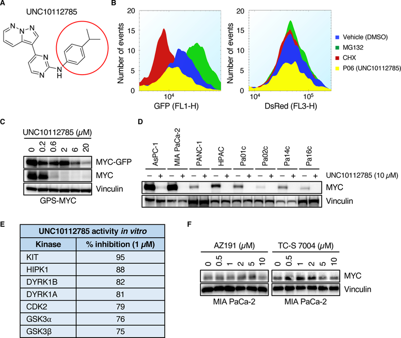
(A) Chemical structure of UNC10112785. The red circle indicates the position at which different analogs were synthesized. (B) GPS-MYC cells were treated with 20 μM UNC10112785 for 6 hours and EGFP and DsRed intensity was measured by flow cytometry. Data from the GPS-MYC screen. (C) GPS-MYC cells were treated with UNC10112785 for 6 hours and EGFP-MYC and MYC levels were measured by immunoblotting. (D) PDAC cells were treated with UNC10112785 and MYC protein levels were measured by immunoblot. (E) Kinase selectivity of UNC10112785 as described previously (29). (F) MIA PaCa-2 cells were treated for 6 hours with the indicated compounds and MYC protein levels were measured by immunoblot. All data are representative of at least three independent experiments.