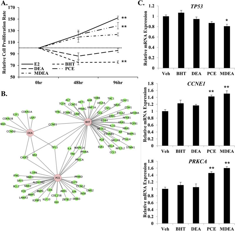
FIGURE 10. Chemical exposure and cell proliferation in Ishikawa cells.
(A) Cell proliferation assays were performed on Ishikawa cells treated for 48 or 96 hrs with 10 nM E2, 100 nM BHT, 100 nM DEA, 100 nM PCE, or 100 nM MDEA. Numbers of cells were normalized to the vehicle-treated plates for that time-point. (B) Genes regulated by BHT, DEA, or PCE that are involved in proliferation of epithelial cells were determined by cross-referencing chemical-associated genes from the Comparative Toxicogenomics Database with gene functions from Ingenuity Pathway Analysis. Gene network was created using Cytoscape. (C) Expression of tumor protein 53 (TP53), cyclin E1 (CCNE1), and protein kinase C alpha (PRKCA) were evaluated by qRT-PCR in Ishikawa cells treated for 6 hr with vehicle, 100 nM BHT, 100 nM DEA, 100 nM PCE, or 100 nM MDEA. Values were normalized to the reference gene peptidylprolyl isomerase B (PPIB) and set relative to vehicle. Bar graphs represent means of at least five biological replicates ± SEM. *p<0.05, **p<0.01 as determined by ANOVA with Tukey’s post-hoc analysis.

