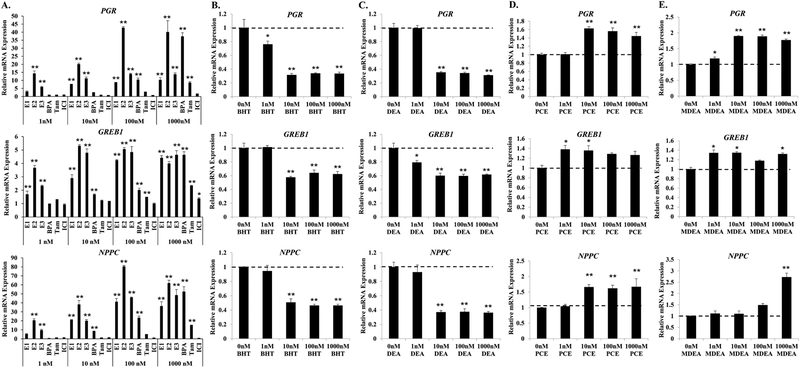
FIGURE 3. BHT, DEA, PCE, and MDEA exposure and transcript levels of endogenous estrogen-responsive genes in Ishikawa cells.
Transcript levels of the progesterone receptor (PGR), growth regulation by estrogen in breast cancer 1 (GREB1), and natriuretic peptide C (NPPC) mRNA were evaluated by qRT-PCR in Ishikawa cells treated for 6 hr with vehicle (0 nM) or 1, 10, 100, or 1000 nM (A) positive and negative reference chemicals, (B) BHT, (C) DEA, (D) PCE, or (E) MDEA. Values were normalized to the reference gene peptidylprolyl isomerase B (PPIB) and set relative to vehicle. Bar graphs represent means of at least five biological replicates ± SEM. *p<0.05, **p<0.01 as determined by ANOVA with Tukey’s post-hoc analysis when compared to vehicle or 0 nM treatment.

