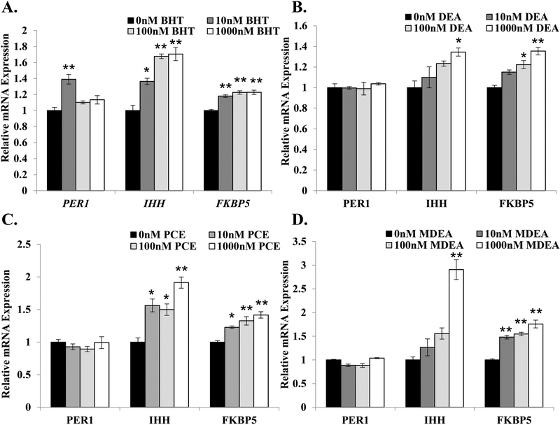
FIGURE 4. BHT, DEA, PCE, and MDEA exposure and transcript levels of endogenous glucocorticoid- and progesterone-responsive genes in Ishikawa cells.
Levels of periodic circadian regulator 1 (PER1), Indian hedgehog (IHH), and FK506 binding protein 5 (FKBP5) transcripts were evaluated by qRT-PCR in Ishikawa cells treated for 6 hr with vehicle (0 nM) or 1, 10, 100, or 1000 nM (A) BHT, (B) DEA, (C) PCE, or (D) MDEA. Values were normalized to the reference gene peptidylprolyl isomerase B (PPIB) and set relative to vehicle. Bar graphs represent means of at least five biological replicates ± SEM. *p<0.05, **p<0.01 as determined by ANOVA with Tukey’s post-hoc analysis.

