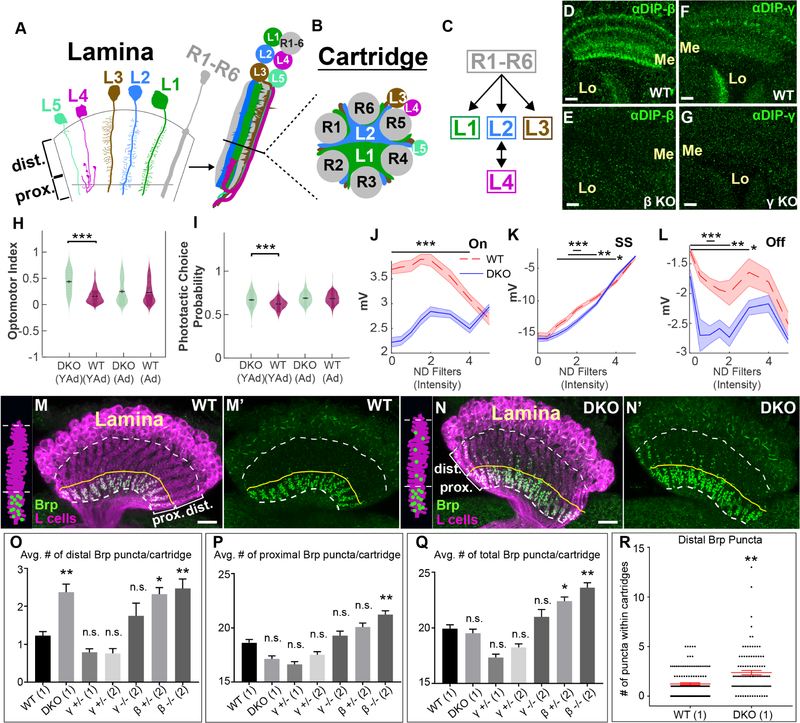
Figure 1. DIP proteins are required for visual function and proper synaptic connectivity.
(See also Figure S1)(A-C) Cellular and synaptic organization of the lamina.
(D-G) Confocal images showing DIP-β (D and E) or DIP-γ (F and G) immunolabeling (green) in the medulla (Me) and lobula (Lo) neuropils at 40–48 hours after puparium formation (h APF). Scale bar = 10μm.
(D and F) In wild type flies DIPs-β and γ are expressed in the medulla and lobula. n=6 brains and n=7 brains respectively.
(E and G) The expression of DIPs-β and γ in the medulla and lobula is severely reduced in flies homozygous for DIP-β (DIP-β1−95) (n=6 brains) or γ (DIP-γ1−67) null mutations (n=5 brains), respectively.
(H) Young adult (YAd; 1–2 day old) DKO flies (n=205) show enhanced tracking of a rotating optomotor stimuli compared to control flies (n=218). Adult flies (Ad; 13–15 days) showed no difference between DKO (n=163) and CTL (n=233) flies.
(I) Young adult (YAd; 1–2 day old) DKO flies (n=105) show an enhanced preference towards the lit arm of a phototactic choice y-maze compared to control flies (n=198). Adult flies (Ad; 13–15 days) showed no difference between DKO (n=168) and CTL (n=232) flies.
(J-L) ERG responses of 1–2 day old DKO (n=9) and control (n=7) flies by intensity for the On transient
(J), steady state response (K) and Off transient (L) components of the ERG in response to a 0.6 second flash of light (See figure S1F). Bars indicate statistical significance at the indicated intensities. Plots in J-L indicate mean of all measurements, shaded areas indicate the SEM).
(M-N’) Confocal images (longitudinal plane of the lamina cartridges) showing the distribution of Brp (green-smFPV5) expressed in L cells (magenta-LexAop-myr-tdTOM) in the laminas of wild type or DKO flies. Scale bar = 10μm. White dotted lines indicate the lamina neuropil. The yellow lines show the boundary between the distal and proximal lamina. We observed batch differences in the amount of Brp background signal in lamina neuron cell bodies (N, N’, O, O’).(M and M’) In wild type flies (n= 9 brains), Brp is restricted to the proximal lamina where L2 and L4 neurons are known to form synapses. (N and N’) In DKO flies (n= 7 brains), Brp is still localized to the proximal lamina, but ectopic Brp puncta are present in the distal lamina.
(O-Q) The average number of Brp puncta in the distal (O) or proximal (P) halves of lamina cartridges from different genotypes are shown. (Q) Indicates the average total number of Brp puncta within lamina cartridges. Results are from two different experiments (1/2). Statistical significance was established with respect to wild type flies (see methods section for a detailed description of statistical analyses). Data are represented as a mean +/− SEM.
(R) Shows the number of distal Brp puncta within individual cartridges scored in wild type and DKO flies. Each star indicates a cartridge. +/− SEM is shown in red. (Statistical significance- *<.05, **<.005, ***<.0005)

