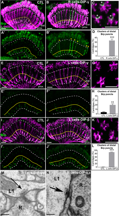
Figure 4. DIP mis-expression promotes synapse formation with Dpr-expressing lamina neurons.
(See also Figures S4 and S5, and Table S1)
(A-B’) Confocal images (longitudinal plane of lamina cartridges, 1–2 day old adults) showing the distribution of Brp (green-smFPV5) expressed in L cells (magenta-LexAop-myr-tdTOM) in the laminas of control (UAS-DIP-γ) flies or flies expressing DIP-γ in R cells (UAS-DIP-γ and GMR-GAL4). The white dotted line outlines the lamina and the yellow line separates the proximal (prox.) and distal lamina (dist.). Scale bar = 10μm.
(A and A’) Brp is restricted to the proximal lamina in control flies.
(B and B’) Streams of ectopic Brp puncta are detected throughout lamina cartridges (yellow stars in B’) when mis-expressing DIP-γ in R cells.
(C) A confocal image of a cross section through the lamina of a fly mis-expressing DIP-γ in R cells. Asterisks indicate the presumed positions of photoreceptor axons. Scale bar = 5μm.
(D) Quantification of the percentage of cartridges containing clusters of Brp puncta in the distal lamina in control flies (n= 11) and flies mis-expressing DIP-γ in R cells (n= 11). Clusters were defined as ≥3 consecutive z-stack slices containing distal Brp puncta. Only unmerged cartridges were considered in the quantification (n=25 cartridges/brain). Data are represented as a mean +/− SEM.
(E-F’) Mis-expression of DIP-γ in L cells (27G05-GAL4). Confocal images show the distribution of Brp (green-smFPV5) expressed in L cells (magenta-LexAop-myr-tdTOM) in the laminas of control flies (27G05-GAL4 alone) or experimental flies (27G05-GAL4 and UAS-DIP-γ). The region above the yellow line delineates the distal lamina (dist.). Scale bar = 10μm.
(E and E’) Brp is localized to the proximal lamina in control (27G05-GAL4) flies, occasionally with some puncta in the distal lamina. n=5 brains.
(F and F’) L cells form ectopic synapses in the distal regions of lamina cartridges (yellow stars in F’) upon mis-expression of DIP-γ in L cells. n= 5 brains.
(G) A confocal image of a cross section through the lamina of a fly mis-expressing DIP-γ in L cells. Scale bar = 5μm.
(H) Quantification of percentage of cartridges containing clusters of Brp puncta in the distal lamina in control flies (n=5) and flies mis-expressing DIP-γ in L cells (n=5). Clusters were defined as ≥5 distal Brp puncta within 5 consecutive z-stack slices of each cartridge (n=25 cartridges/brain). Data are represented as a mean +/− SEM.
(I and I’) Brp is localized to the proximal lamina in control (UAS-DIP-β) flies. n=5 brains.
(J and J’) In flies mis-expressing DIP-β in L cells (27G05-GAL4), ectopic synapses are present throughout lamina cartridges. n=7 brains.
(K) A confocal image of a cross section through the lamina of a fly mis-expressing DIP-β in L cells. Scale bar = 5μm.
(L) Quantification of percentage of cartridges containing clusters of Brp puncta in the distal lamina in control flies (n=5) and flies mis-expressing DIP-β in L cells (n=5). Clusters were defined as ≥5 distal Brp puncta within 5 consecutive z-stack slices of each cartridge (n=25 cartridges/brain). Data are represented as a mean +/− SEM.
(M and N) Putative L1-R cell synapses in flies mis-expressing DIPs-γ and ε in R cells identified by EM.
Scale bar = 200nm.
(Statistical significance- *<.05, **<.005, ***<.0005)

