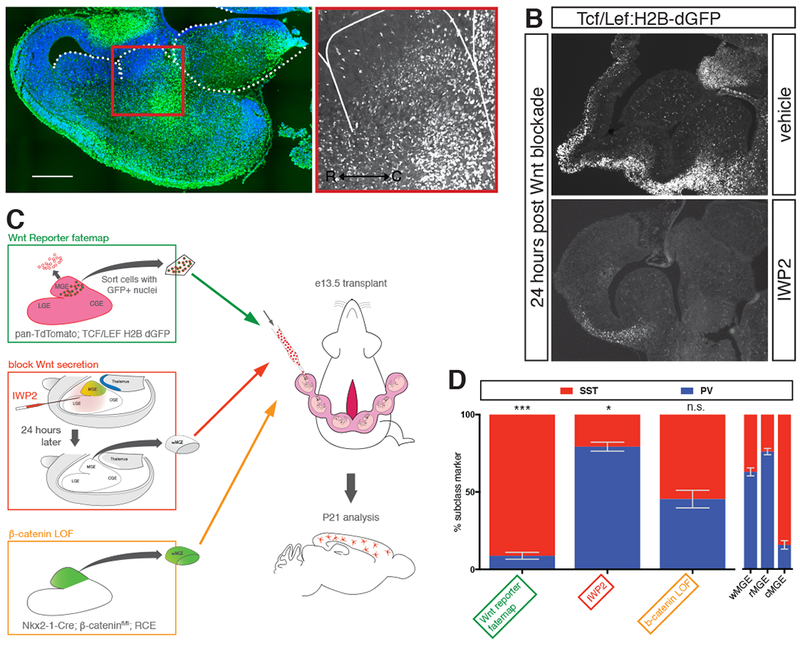
Figure 3. Exploring the role of Wnt signaling in determining cortical interneuron subtype identity.

(A) Transverse section of e13.5 mouse brain in Tcf/Lef:H2B-dGFP reporter mouse. Nuclear GFP is enriched in caudal portion of MGE. (B) Parasagittal section of Tcf/Lef:H2B-dGFP reporter mouse at e13.5 showing regions of Wnt-responsiveness. 24 hours after intraventricular injection of vehicle did not affect GFP signal. 24 hours following intraventricular injection of IWP2 strongly reduced GFP signal. (C) Schematic diagram depicting the different transplant studies performed to examine the role of Wnt in interneuron specification. Green box shows pan-RFP+ cells sorted for eGFP+ nuclei indicative of Wnt responsiveness. Red box shows intraventricular injection of IWP2 at e12.5, followed by dissection and transplant of wMGE 24 hours post-injection. Orange box shows genetic removal of b-catenin and simultaneous eGFP+ fate mapping of MGE prior to dissection and transplantation. (D) P21 analysis of studies transplanting Tcf/Lef:H2B-dGFP+ MGE cells, blocking Wnt secretion prior to transplantation, or genetic removal of b-catenin in transplanted MGE. W-, r- and cMGE transplant results on right for comparison. Error bars standard error of the mean (* denotes p<0.05; ** denotes p<0.01; *** denotes p<0.001).
