
Figure 4. Identifying components of non-canonical Wnt and Ryk signaling in the embryonic MGE.
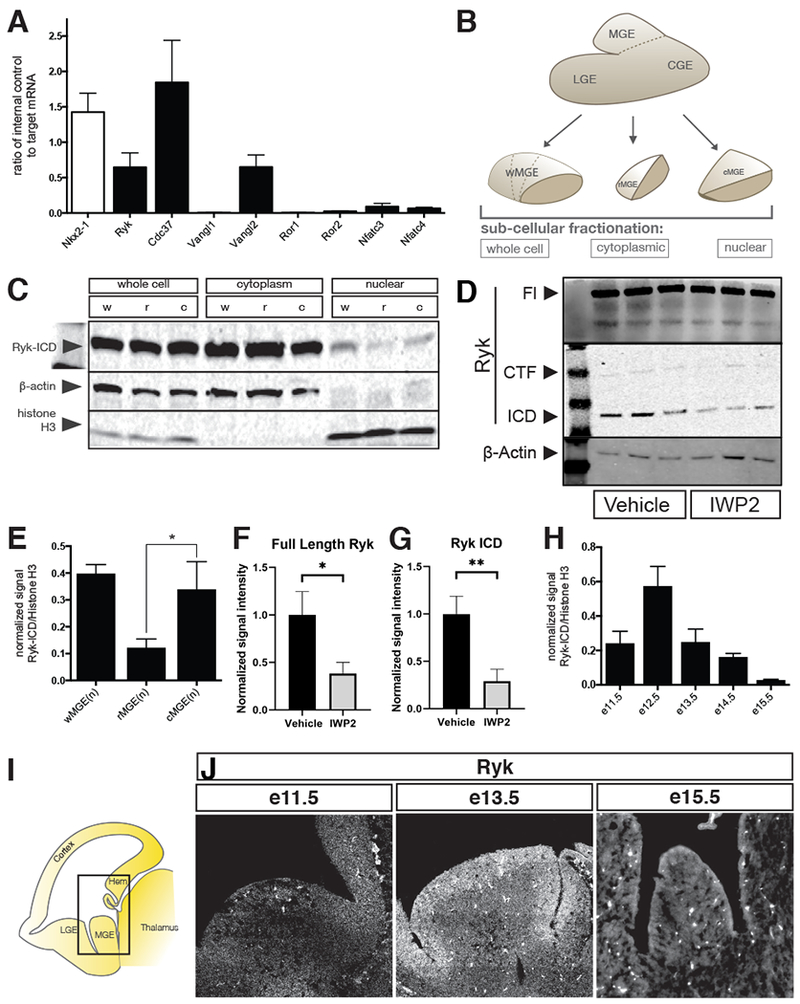
(A) Quantitative PCR of e12.5 MGE for non-canonical Wnt signaling components, Nkx2-1 shown as a positive control. (B) Schematic depiction of various MGE samples loaded for western blot in C. See also Supplemental figure 3. (C) Biochemical fractionations of w-, r- and cMGE into whole cell, cytoplasmic and nuclear components were analyzed by Western blot for Ryk, b-actin and histone H3 (latter two act as loading controls). (D) FL Ryk and Ryk ICD fragments are decreased in the cytoplasm of MGE cells 24hrs after IWP2 treatment. (E) Quantitation of blot in C: nuclear Ryk intracellular domain (Ryk-ICD) present in w-, r- and cMGE samples. (F,G) Quantification of blot shown in D, normalized to loading control and average signal intensity of the vehicle treated bands. (H) wMGE nuclear fractions at various embryonic time points showing that Ryk signaling is dynamic over time. (I) Schematic of parasaggital sections of 13.5 mouse brain. Equivalent areas at e11.5, e13.5 and e15.5 show decreasing levels of Ryk receptor over developmental time by (J) in situ hybridization for Ryk.