
Figure 5. Ryk loss- and gain-of-function and its effects on cortical interneuron specification.
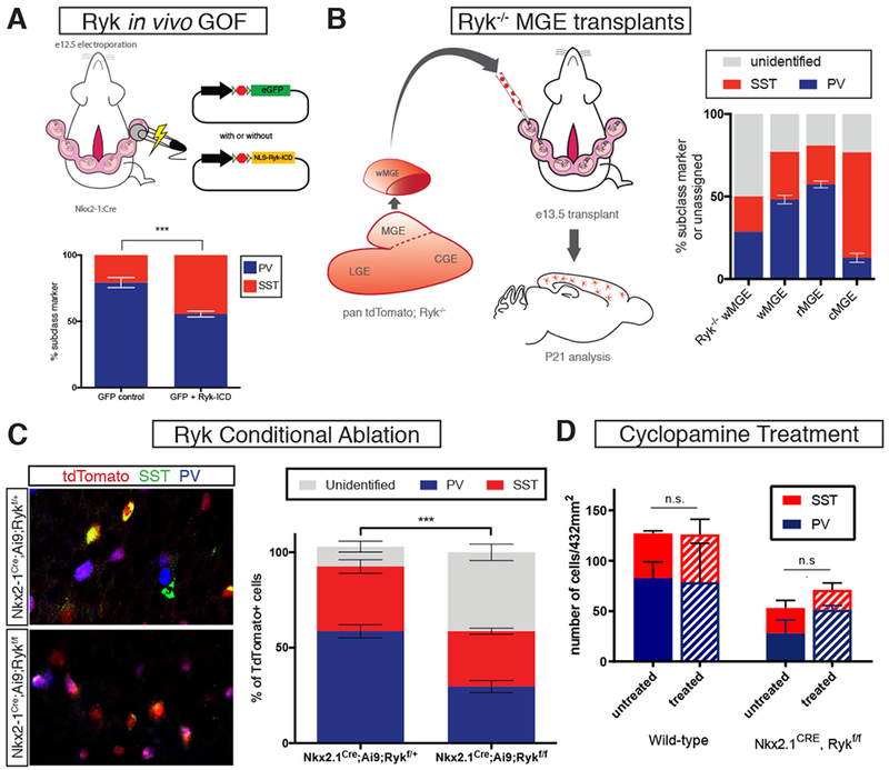
(A) Left, schematic diagram of electroporation paradigm for Ryk ICD gain-of-function in MGE progenitors. Right, analysis of PV+ and SST+ interneurons at P21 with control (GFP alone) or Ryk ICD gain-of-function (NLS-Ryk-ICD) plasmid electroporation. Ryk-ICD GOF resulted in a significant increase in SST+ and a decrease in PV+ electroporated interneurons. (B) Schematic diagram showing Ryk−/− MGE transplant study. (B) P21 analysis revealed that Ryk−/− MGE transplants contain a large percentage of unidentified cortical interneurons; wild type w-, r-, and cMGE transplant results for comparison. (C) Conditional genetic ablation of Ryk and simultaneous labeling with tdTomato in the MGE using Nkx2-1Cre. Left top, representative image of Rykf/+, left bottom, representative image of Rykf/f. tdTomato in red, somatostatin in green, parvalbumin in blue. Asterisks denote unidentified tdTomato+ cells. Right, quantification of unidentified, somatostatin+ and parvalbumin+ tdTomato+ cells for each group. Error bars standard error of the mean (* denotes p<0.05; ** denotes p<0.01; *** denote p<0.001). (D) Treatment of Nkx2.1Cre; Rykf/f or WT embryos at e12.5 with cyclopamine has no significant nor additive effect on the numbers of PV+ and SST+ interneurons observed in adults.