Fig. 2.
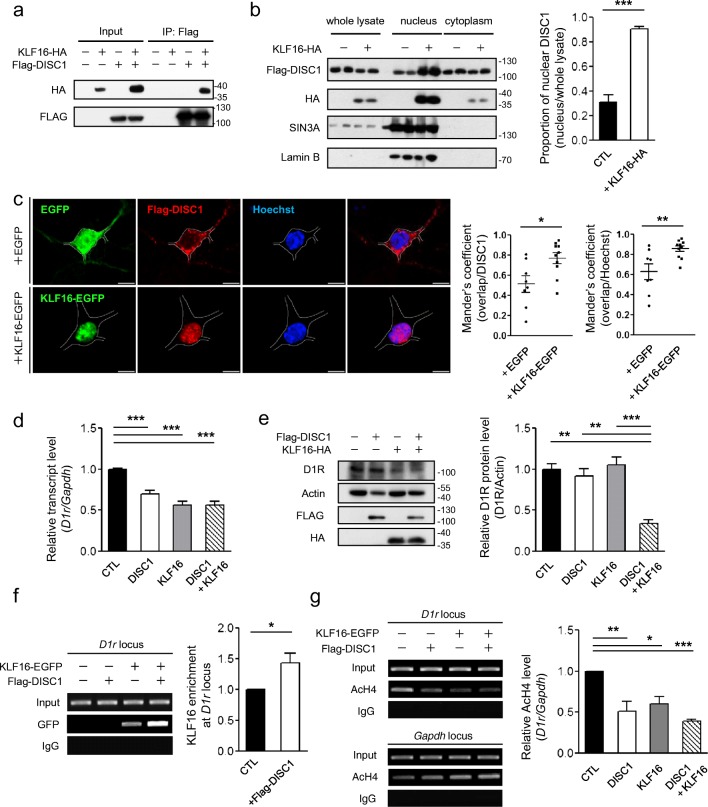
Translocation of DISC1 into the nucleus via interaction with KLF16. a CoIP of KLF16-HA with Flag-DISC1 using anti-Flag antibody from differentiated CAD cell lysates. b Increased DISC1 protein level in the nucleus-enriched fractions of differentiated CAD cell lysates (n = 3). c Confocal images of Flag-DISC1 (red) and the Hoechst 33342 (blue) upon EGFP or KLF16-EGFP (green) overexpression. Mander’s overlap coefficients between Flag-DISC1 and Hoechst were calculated by the Cellsens software (Olympus) (n = 8 for EGFP, n = 10 for KLF16-EGFP, E15.5 mouse striatal cultured neurons at DIV10). The white line outlines the neuronal soma. The scale bar represents 5 μm. d Reduced transcript level of D1r upon DISC1, KLF16, and their combined overexpression in differentiated CAD cells (n = 8). e Reduced endogenous D1R protein level upon DISC1 and KLF16 co-expression (n = 3). f Enhanced ChIP-PCR signal of KLF16-EGFP at the D1r locus in DISC1-cotransfected CAD cells (n = 5). Anti-GFP antibody was used for IP. g Reduced acetylated H4 level at the D1r locus upon DISC1, KLF16, and their combined overexpression in differentiated CAD cells (n = 4). Anti-AcH4 (K5, K8, K12, and K16) antibody was used for IP. Data represent mean ± SEM. *P < 0.05; **P < 0.01; ***P < 0.001; two-tailed unpaired t test (b, c), paired t test (f); one-way ANOVA with post-hoc Tukey test (d, e, g)

