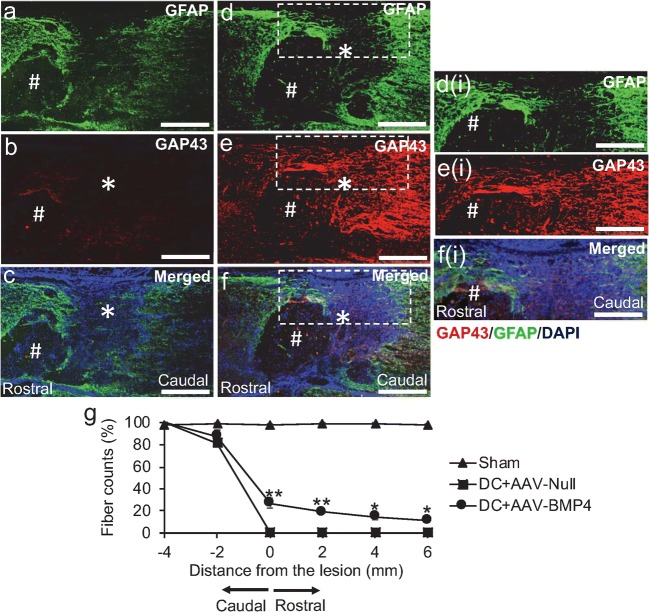
Fig. 4.
BMP4 promotes axon regeneration in an in vivo rat model that cavitates. a–c Treatment with DC + AAV-Null leads to cavity (#) formation at the lesion site (*) with no evidence of axon regeneration. d–f AAV-BMP4 enhances DC axon regeneration despite the presence of cavities (#), detected by GAP43+ staining (red) with GFAP+ astrocytes (green) also infiltrating the lesion site (*). c, f Merged images to show GFAP (green) and GAP43 (red) double staining with DAPI+ nuclei (blue). Inset d(i), e(i) and f(i) = higher power views of the boxed regions in d–f, respectively. g Quantification of DC axon regeneration at the lesion site, rostral and caudal to the lesion. *** = P < 0.0001, ANOVA. ** = P < 0.001; * = P < 0.05, ANOVA. Scale bars in a–f and d(i)–f(i) = 100 μm

