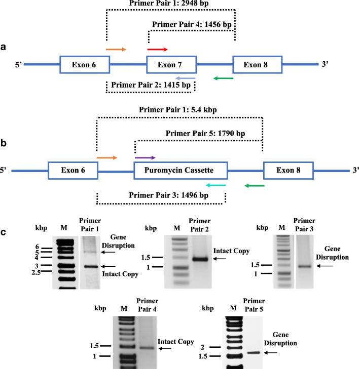
Fig. 2.
Analysis of RARα gene disruption. a Schematic drawing of the RARα wild type gene structure showing the region between exon 6 and exon 8. b RARα gene structure after CISPR-Cas9-mediated replacement of exon 7 by the puromycin expression cassette. The diagnostic PCR amplicons produced using different pairs of primers are shown. Primer pairs 2 and 4 are specific for the wild type RARα gene while primer pairs 3 and 5 detect the insertion of the puromycin expression cassette. Primer pair 1 spans the region containing exon 7 and produces a longer product when the puromycin expression cassette has been inserted. c Characterisation of a hemizygous RARα± cell line. Genomic DNA isolated from a clonal SH-SY5Y cell line was analysed using the primer combinations described above, and amplicons were analysed by 1% agarose gel electrophoresis. Both puromycin cassette and exon 7 were detected in the clonal SH-SY5Y cells which indicate that only one copy of RARα was knocked out. M is a DNA size standard

