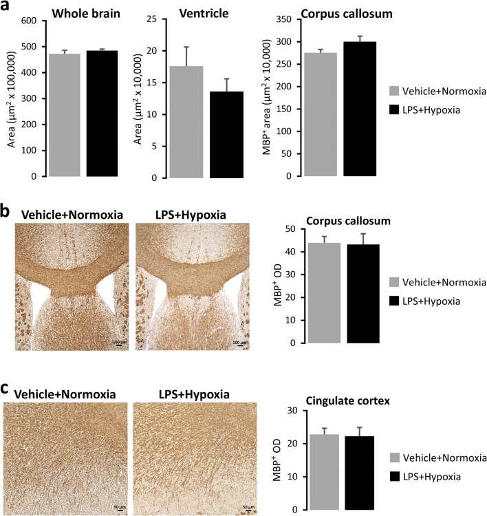
Fig. 6.
Immunohistological analysis of the brains of 50-day-old male mice. a Area measurements (μm2) of whole brain, ventricles and corpus callosum. b, c Representative images of corpus callosum (b) and cingulate cortex (c). MBP positive staining performed on 10-μm-thick cryosections from P50 mice neonatally exposed to Vehicle + Normoxia or LPS + Hypoxia. Bar graphs show means ± SEM of MBP positive optical density (OD) (n = 6/group). Scale bars: 100 μm (b), 50 μm (c). Abbreviation: LPS lipopolysaccharide

