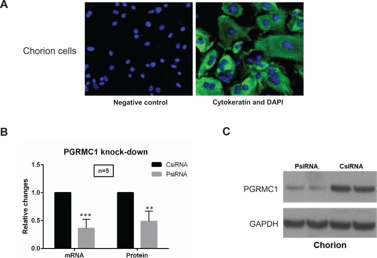
Figure 1.
A, Immunofluorescence staining of chorion cells. Anti-human cytokeratin antibody was used for staining to distinguish chorion cell cultures (green) from mesenchymal type of cells along with observation of cell morphology. DAPI counterstaining was performed to visualize nuclei (blue). Images of cells were captured using digital camera interfaced with a fluorescence microscope. Magnification is ×40. B, C, Progesterone receptor membrane component 1 (PGRMC1) messenger RNA (mRNA) and protein knockdown in chorion cells. PsiRNA is for PGRMC1 small-interfering RNA (siRNA)-transfected group; CsiRNA is for control siRNA-transfected group. qPCR (B) and Western blot (C) were performed to confirm the knockdown of PGRMC1 mRNA (in 24 hours) and protein expression (in 72 hours), respectively. Representative Western blot and densitometry data are presented in (C).

