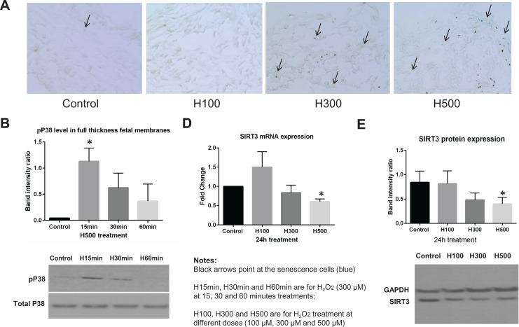
Figure 4.
Hydrogen peroxide (H2O2)-induced cell senescence, p38 mitogen-activated protein kinase (MAPK) phosphorylation, and downregulated sirtuin 3 (SIRT3) expression in full-thickness fetal membrane explants. A, Senescence cell histochemical staining of full-thickness fetal membrane explants treated with 0, 100, 300, and 500 μM H2O2 for 24 hours. SA-β-Gal-positive cells are blue in representative images. B, The proportion of SA-β-Gal-positive cells was summarized in a bar graph. C, Representative Western blot image to demonstrate the p38 MAPK phosphorylation under 500 μM H2O2 treatment at 15, 30, and 60 minutes. Densitometry analysis was summarized in a bar graph (N = 6). D, SIRT3 mRNA expression level in fold change calculated using the ΔΔCt method after normalization (24-hour treatments, N = 6). E, Representative Western blot image to demonstrate SIRT3 protein expression and densitometry analysis was summarized in a bar graph (24-hour treatments, N = 6).

